Mr William Flannery BM BS FRCS(ORL-HNS)
Combined Approach Tympanoplasty (CAT)
Introduction
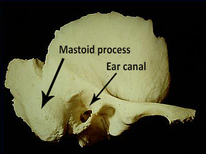
Combined approach tympanoplasty (CAT) is a surgical treatment for chronic ear infections which do not respond to medication and microsuction.There are several approaches for operations on the temporal bone (see photograph). One of the most common after myringoplasty is CAT. The operation is often done in several stages depending on the findings at the first (stage I) procedure.
Overview of a CAT
Again looking at the photograph, working from left-to-right, one approach is made by drilling off the cortex of the mastoid process - the prominence behind the ear - and forming a small mastoidectomy cavity and a second is made by lifting a flap of skin in the ear canal to get down to the ear drum. There is therefore a combined approach to the middle ear structures.FAQs
-
What does 'tympanoplasty' mean?
In chronic middle ear infections, the ear drum (tympanum) often has to be repaired and sometimes the hearing can be improved with grafting. Operating on the drum and the bones of hearing is called tympanoplasty.
-
What is a mastoid cavity?
There is large prominence behind the ear called the mastoid process. In mastoidectomy, a small part of the prominence near the groove where the ear joins the head is drilled out to remove the infected air cells and gain access to the middle ear.
-
Nerve damage during the operation
Damage to the facial nerve (facial paralysis) and chorda tympani (disrupted taste) is possible. I have not had a facial nerve palsy as complication in cases where the bony covering of the nerve was intact pre-op. Where the infection has exposed the facial nerve, I have had two nerve palsies caused by the local anaesthetic which resolved spontaneously. The chorda tympani is often divided deliberately during a CAT. If done cleanly it does not result in disturbance of taste since there is plenty of cross innervation from the opposite side.
-
Surgical incisions
There are two incisions in a CAT. One is hidden behind the ear. The scar can only be seen with very close inspection. The other incision is down the ear canal. It cannot be seen without a microscope and heals without a scar.
-
Healing within the middle ear
In a CAT, the object is to remove trapped skin and inflamed lining from the middle ear and allow normal, surrounding mucosa to grow over the exposed bone. In non-cholesteatoma cases (inflammation/no skin), if the ear settles down clinically there is no need for further procedures/CAT stages. If there is skin trapped or the inflammation does not settle then a second procedure is required.
-
Will the hearing be worse?
The infection process itself often damages the bones of hearing causing a deafness of between 25 to 60 dB. So it is common before the operation to have some degree of hearing loss. The middle bone of hearing called the incus is often covered in drum and needs to be removed. This produces a permanent hearing loss. The magnitude of the loss varies considerably from one case to another but theorectically the worse it can be is 60 dB. This is eminently aidable.
Because there is inevitably some loss of hearing, I make every effort over a prolonged period to try to eradicate the infection in clinic and only resort to surgery when I cannot make any progress.
-
Other details
A stage I CAT operation lasts around 3.5 hours under general anaesthetic. I apply a pressure bandage at the end around the head which stays on overnight. I use dissolvable sutures which do not need removing by the GP. Dressings are left in the ear canal and removed in clinic at the 3 week follow up.